News

A group of extremely rare composite lymphomas reported in American Journal of Surgical Pathology by Yuhua Huang MD.PhD

June 21, 2022

There are many histological subtypes of lymphoma, and the pathological diagnosis of lymphoma is difficult and challenging. Accurate diagnosis and typing are of great significance to the precise treatment of lymphoma patients and to improve the curative rate. In rare cases, two different histological types of lymphoma can occur simultaneously at the same anatomical site, which is called composite lymphoma. At present, little is known about the pathogenesis and clinicopathological features of composite lymphoma.
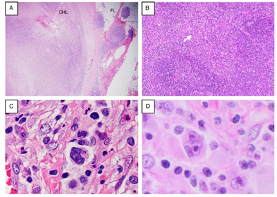
 
Figure 1. Morphologic patterns of composite classic Hodgkin lymphoma and follicular lymphoma
A, in this case the CHL (left) and FL (right) occupy separate areas of the lymph node. B, in this case CHL (arrow) is present in the interfollicular regions between nodules of FL. C and D, Hodgkin/Reed-Sternberg cells in a background of mixed inflammatory cells.

In June, 2022, Yuhua Huang MD.PhD, from the Dept. of Pathology, at Sun Yat-sen University cancer center, reported 22 cases of extremely rare composite lymphoma (composite classical Hodgkin lymphoma and follicular lymphoma, CHLFL) in the American Journal of Surgical Pathology - a top journal in the field of clinical pathology. The clinicopathological features of this tumor were systemically analyzed. To date, it is the largest report on this rare cancer. Interestingly, all three cases investigated by cytogenetic methods for a clonal relationship between the CHL and FL components were clonally related, suggesting that the CHL and FL components may share a common progenitor B-cell, which is likely a mutated germinal center B-cell. This finding provides a new idea for pathogenic study of this rare tumor.
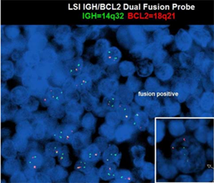
 

Figure 2. Both the follicular lymphoma cells and Hodgkin/ Reed-Sternberg cells (inset, arrow) show IGH/BCL2 fusion (yellow signal) by FISH (green signal IGH, red signal BCL2).

Acknowledgement: this study was guided and supported by Ji Yuan, MD, PhD, the corresponding author of the article, from the Mayo Clinic in the United States. For the original publication, click here.

Yuhua Huang MD. PhD, is currently an associate chief physician in the Department of Pathology, Sun Yat-sen University Cancer Center, Guangzhou, China. He specializes in lymphoid pathology and is one of co-authors of WHO Classification of Tumours of Haematopoietic and Lymphoid Tissues (5th Edition). Dr. Huang was a visiting scholar at the Prince of Wales hospital, the Chinese University of Hong Kong and University of Nebraska Medical Center (UNMC), USA. He is the committee member of the Lymphoma Group, Pathology Branch of Chinese anti-cancer association (CACA) and CCPI. He was also the recipient of the "Outstanding young pathologists" branch of the Chinese Medical Association in 2021. Dr. Huang’s primary research interests include the pathological and transnational research on EBV-associated malignancy, including lymphoma and EBV-associated cholangiocarcinoma. He has published more than 40 peer-reviewed papers, including papers in renowned journals such as the Journal of Hepatology, the American Journal of Surgical Pathology, the International Journal of Cancer, and Histopathology.

Where are we | Contact us|

Copyright:Sun Yat-sen University Cancer Center Designed by Wanhu.